This article was first published in the University of Illinois Urbana-Champaign newsroom. Read the full story here.
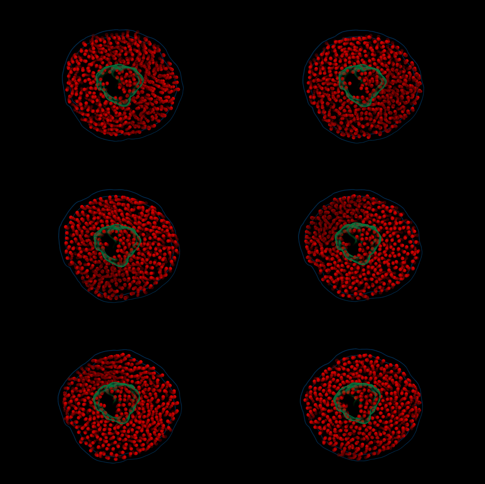
Researchers at Georgia Institute of Technology and the University of Illinois Urbana-Champaign have developed a first-of-its-kind technique called electron videography to capture moving images at the molecular scale. In the first demonstration of the technique, the team took a microscopic moving picture of the delicate dance between proteins and lipids found in cell membranes. The study, “Electron videography of a lipid–protein tango” was published last week in the journal Science Advances.
"This is the first time we are looking at a protein on an individual scale and haven't frozen it or tagged it," says Aditi Das, a corresponding author and associate professor in the School of Chemistry and Biochemistry at Georgia Tech.
Electron microscopy techniques image at the molecular or atomic scale, yielding detailed, nanometer-scale pictures. However, they often rely on samples that have been frozen or fixed in place, leaving scientists to try to infer how molecules move and interact — like trying to map the choreography of a dance sequence from a single frame of film.
"Usually, we have to crystalize or freeze a protein, which poses challenges in capturing high-resolution images of flexible proteins. Alternately, some techniques use a molecular tag that we track, rather than watching the protein itself,” Das says. “In this study we are seeing the protein as it is, behaving how it does in a liquid environment, and seeing how lipids and proteins interact with each other."
The technique can be used to study the dynamics of other biomolecules, breaking free of constraints that have limited microscopy to still images of fixed molecules. In this study, the team examined nanoscale discs of lipid membranes and how they interacted with proteins normally found on the surface of or embedded in cell membranes.
These membrane proteins are significant for medical treatments, and are involved in processes including muscle contraction, brain function, and immune system functions. Moving forward, the researchers plan to use their electron videography technique to study other types of membrane proteins and other classes of molecules and nanomaterials.
For More Information Contact
Contact:
Jess-Hunt Ralston
Director of Communications
College of Sciences
Georgia Tech


